Suspected Posterior MI Suspected MI with a non-diagnostic ECG Record leads V7-V9 Correlates with posterior wall MI Left circumflex infarct related artery in all J Am Coll Cardiol 199934748. A posterior wall MI occurs when posterior myocardial tissue now termed inferobasilar usually supplied by the posterior descending artery a branch of the right coronary artery in 80 of.

Heart Anatomy Anatomy And Physiology Ii
Beginning with the arrowhead view depicted as a V the probe is angled caudally to depicted initially the pulmonary artery RVOT is seen as a longitudinal vessel with blood flowing towards the spine anterior to posterior.

. The system enables surgeons to create a soft landing of posterior spinal constructs preventing proximal junctional kyphosis in spinal deformity cases according to a news release. The cervical spine series is a set of radiographs taken to investigate the bony structures of the cervical spine albeit commonly replaced by the CT the cervical spine series is an essential trauma radiograph for all radiographers to understand. Expiration to minimize superimposition of the diaphragm over the upper lumbar spine centering point.
A left posterior fascicular block also known as a left posterior hemiblock LPHB occurs on the ECG when the posterior fascicle of the left bundle branch is no longer able to. Posterior Tilt Hip Biomechanics. On its anterior border and to its right is the SVC and RA and to its left and anterior is the coronary.
As with the posterior and anterior approaches instrumentation corrects the curve and the rotation of the spine. The spinal cord is a long thin tubular structure made up of nervous tissue which extends from the medulla oblongata in the brainstem to the lumbar region of the vertebral columnIt encloses the central canal of the spinal cord which contains cerebrospinal fluidThe brain and spinal cord together make up the central nervous system CNS. The first cases with Implanets Jazz posterior fixation system were completed the devicemaker said Feb.
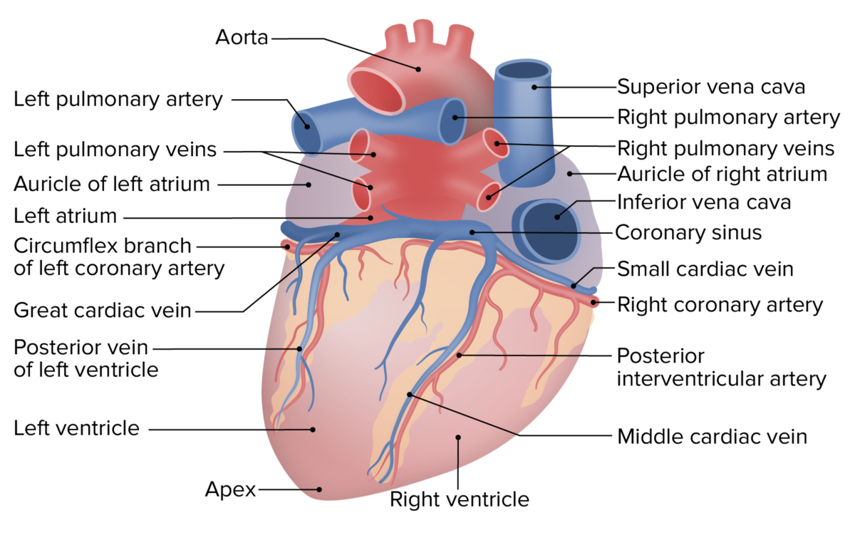
An axial view of the ascending aorta is seen immediately to the right. Aortic valve located between the left ventricle and the ascending aorta aortic orifice. In its typical anatomical orientation the heart has 5 surfaces formed by different internal divisions of the heart.
Posterior scapula line V9. Blood supply to the tibialis posterior muscle is through branches of the posterior tibial artery which stems the popliteal artery. M1 Anterior Neck and Thorax Back and Spinal Cord Larynx Pharynx and Cervical Sympathetic Trunk Superior Mediastinum and Root of Neck Heart Lungs and Ventilation Pathway Posterior Mediastinum Kidneys and Urinary System Abdominal Wall Peritoneum and Intestines Stomach Liver and Spleen Duodenum.
On the superior and inferior surfaces of the lateral masses are facets for articulations with other bones. The left Posterior Fascicular block Lpfb is defined by irregular contraction of heart muscles whereby it travels to the inferior and posterior portion of the left ventricle of the heart and does not conduct electrical impulse transmission from the atrioventricular node. It is the most central of all the chambers but sometimes lies just left of midlene.
Adults with scoliosis and spinal stenosis often also require a decompression procedure in which the roof of the vertebral column is removed at the affected area freeing the nerve from any material that is compressing it prior to. Cervical spine radiographs are indicated for a variety of settings including 1-3. Muscles along the spine Calves.
Tibialis posterior is innervated by the tibial nerve which arises from the L4 and L5 spinal nerves. Because of this all of these diagnoses must be considered for effective treatment of posterior heel pain. In a posterior tilt the upper part of the pelvis is positioned behind the imaginary vertical plumb line or at least as can be the case during exercise is moving in that direction.
Gastrocnemius and soleus The posterior chain also includes muscles in the upper body such as the trapezius latissimus dorsi and rhomboids. The posterior arch instead of a spinous process has a posterior protrusion known as the posterior tubercle. For example a patient with a bone spur of the calcaneus may have bursitis in that area as well.
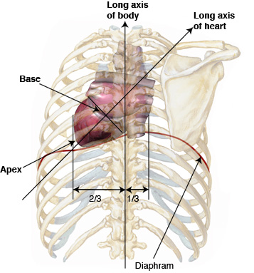
In humans the spinal cord begins at the. The apex of this pyramid pointing in an anterior-inferior direction. Anterior or sternocostal Right ventricle.
Same horizontal plane as V4. The heart has been described by many texts as a pyramid which has fallen over. Superior to inferior normal heart anatomy.
Left border of spine V5-V9. The left atrium is the most posterior of the heart structures with its most immediate posterior relation being the esophagus and then the spine and and aorta. This allows the removal of disk herniation and relieves pressure on spinal nerves but the dissection of spinal muscles and supporting tissues can.
Superiorly to include T12L1. In conventional diskectomy for example the paraspinal muscles are dissected from the posterior aspect of the lumbar spine and portions of the lamina are removed to gain access to the spinal canal. The valve consists of three cusps left right and anterior named by their position in the foetus before the heart undergoes rotation.
Diagnosis of the cause of posterior heel pain can be difficult as it is not uncommon for these diagnoses to coexist. In addition grooves for the vertebral arteries lay just posterior to the superior articular facets located on the lateral masses. The tibial nerve is the larger of the two branches of the sciatic nerve.
Posterior or base. Posterior axillary line V8. The aortic valve consists of three cusps right left and posterior.
As the upper part of the pelvis is pulled backward the bottom part of the pelvis is pulled forward. The level of the iliac crest coronal centering point is directly over the lumbar vertebra which corresponds to the posterior third of the abdomen the central ray is perpendicular to the image receptor collimation.

Heart Anatomy Anatomy And Physiology Ii
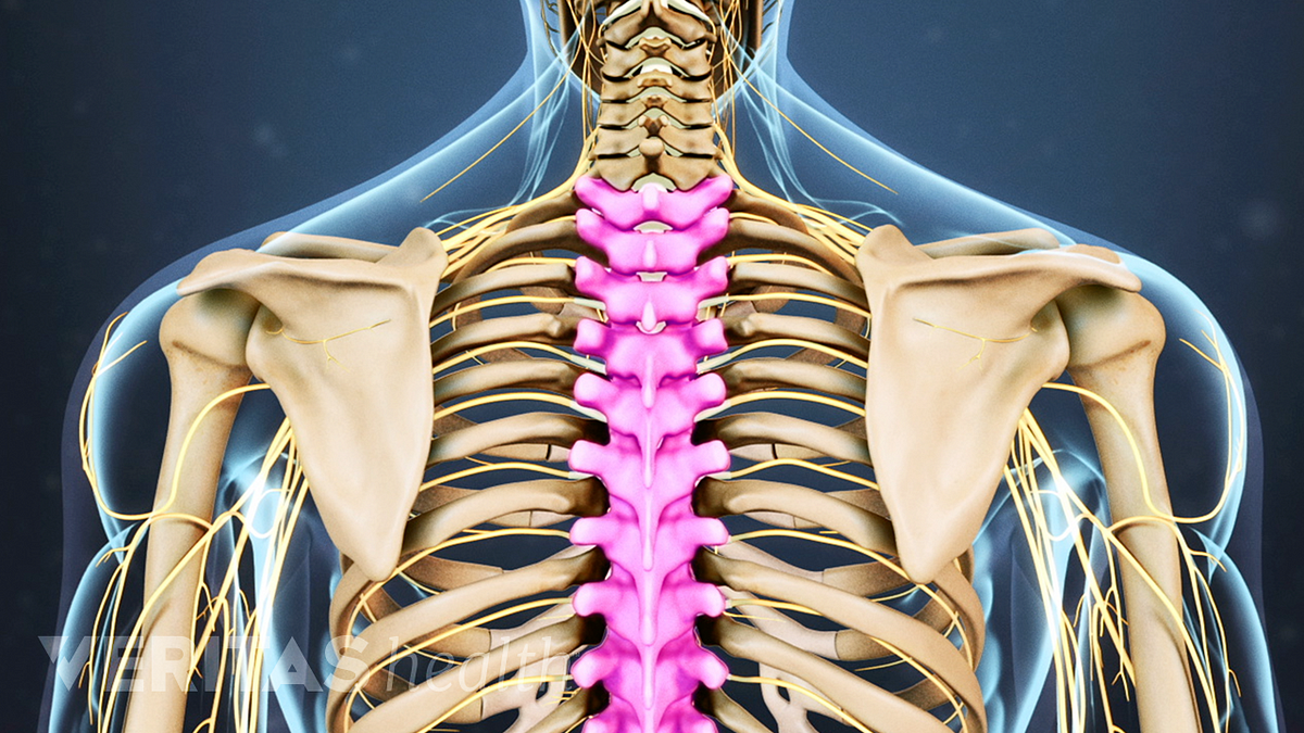
Thoracic Spine Anatomy And Upper Back Pain

Posterior View Of The Organs Within The Abdominal Cavity The Lumbar Download Scientific Diagram
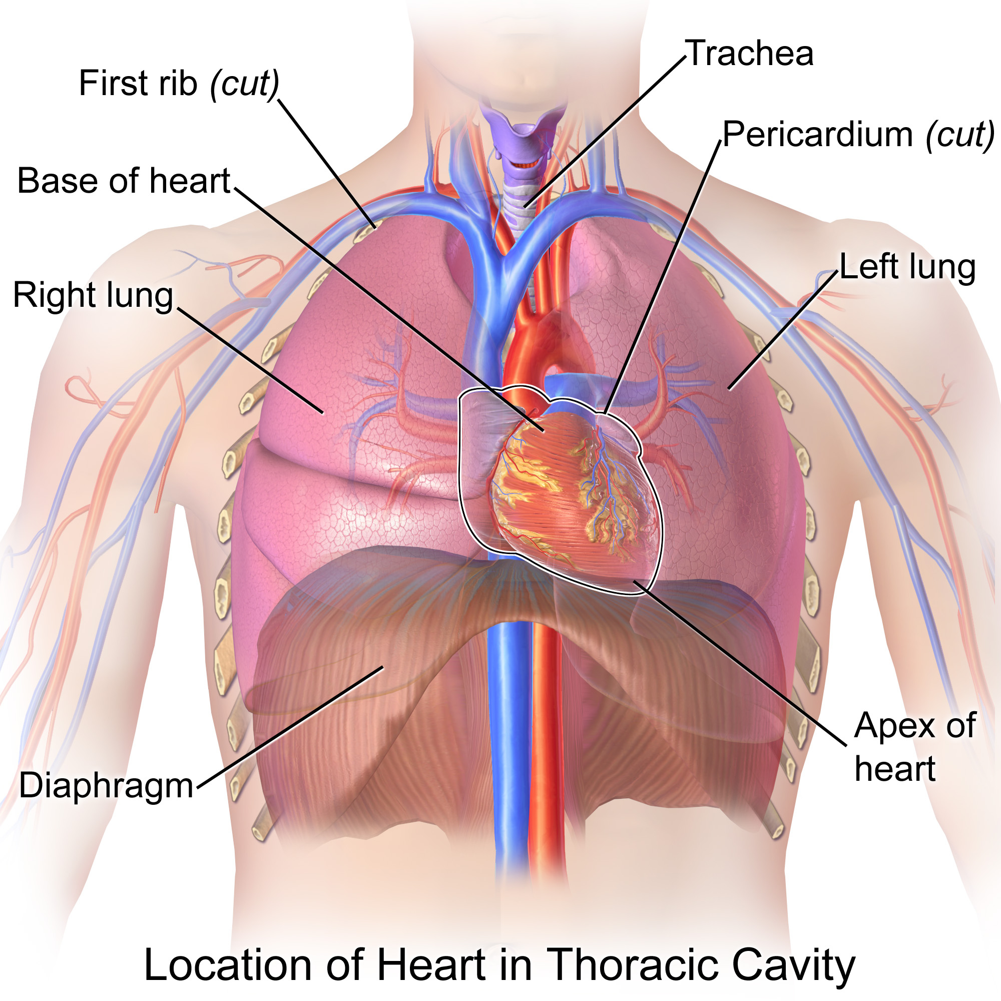
Is The Heart Located Posterior Or Medial To The Lungs Socratic
Heart Anatomy Concise Medical Knowledge

Positioning Of The Heart
Heart Anatomy Anatomy And Physiology Ii
Anatomy Tutorial Posterior Atlas Of Human Cardiac Anatomy
0 comments
Post a Comment